At VASTA, we are naturally skeptical of ‘new’ treatment approaches, especially those that make BIG promises. Our clinicians are current with best practices and are committed to an evidence-based approach.

At the same time, we work hard to stay on the cutting edge of emerging treatment techniques.

Blood Flow Restriction (BFR) Training is one such approach.

Recently, this little known technique has become a popular tool at military hospitals for soldiers who experienced significant limb trauma (e.g. in ‘limb salvage’ wards) and with amputees. Millions of dollars of research has been poured into exploring the utility of BFR for patients with significant limitations (i.e. limited mobility and/or limited ability to bear weight) who need to build muscle.  A very difficult task!

Similarly, our patients with post-operative limitation that prohibits weight bearing or heavy use of the LE for weeks (or even months) end up with dramatic muscle atrophy. Previously thought of as a ‘necessary evil’ to protect the surgical repair… Now, maybe not!

BFR involves restricting the blood flow to a limb during exercise in order to starve the muscle of oxygen, thus causing a number of ‘pro-growth’ chemical and hormonal changes. This allows us to stimulate muscle hypertrophy (growth) with very low intensity & low load exercises.

For any patient requiring a prolonged period of ‘rest’, immobilization or limited weight bearing – we believe BFR training has the potential to be a game changer.

What is Blood Flow Restriction Training?

Blood flow to the limb is significantly reduced, and blood return (flow out of the limb) is completely stopped, for a brief period of time, with the use of a tourniquet. This specially designed tourniquet contains a Doppler (to sense blood flow) and is automated to control blood flow to precisely defined parameters.

Once blood flow is reduced to the limb, the patient performs ~ 4-5 minutes of low intensity resistance training (LIT) exercises. While the load is very light – and thus the strain to the bone, cartilage, ligaments, etc. is minimal – the muscle gets exhausted! This is because it is being forced to work under anaerobic conditioning (without oxygen from fresh blood supply).  Thus, the muscle seems to work very hard, all muscle fiber types are recruited, lactic acid builds up, the muscle glycogen levels become depleted and the exerciser feels as though they have just worked out really hard.

More importantly, the muscle also thinks it worked out really hard; and, the response is similar to high intensity training (HIT) – The muscle grows and gets stronger!

Bottom line – this technology allows us to achieve strength and hypertrophy (size) gains that, under normal circumstances, are possible only under high loads & high exercise intensities.

We can make gains earlier and faster, even when an injury prevents you from exercising.

For those looking for more detail and a review of the extensive research that has been done in this field, read on…

Graph 1

 Graph 2

According to the American College of Sports Medicine (ACSM), building muscle strength and size can be achieved through moderate to high intensities (Donnelly 2009) typically at intensities of 65% > 70% of a ‘1 rep max’.

Unfortunately, many of our injured athletes and post-operative patients can not work at that level of intensity. Worst, patients that require prolonged immobilization and/or restricted weight bearing on a limb suffer severe muscle atrophy.

Low intensity exercise, which is tolerated much better in these folks, does not result in strength gains or muscle hypertrophy.

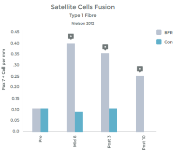
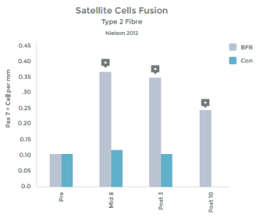
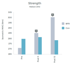
However, exercising at low intensities (LIT) has been shown to result in strength gains and hypertrophy when coupled with BFR!

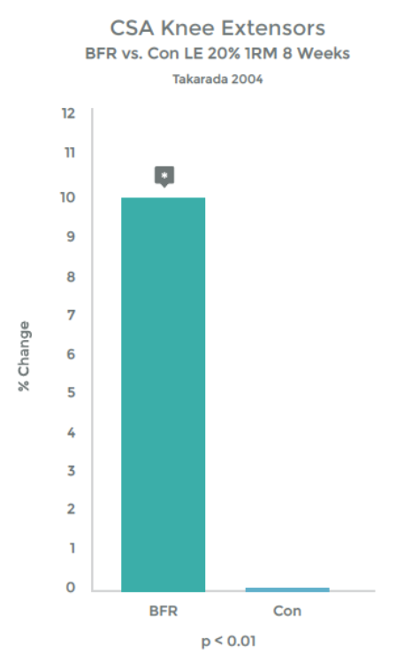
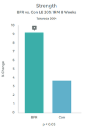
Graph 1 & 2 to left illustrate this finding.

Strength gains & Hypertrophy in the Quadriceps muscle from VERY low load (20% 1RM) exercise over 8 weeks!

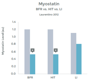
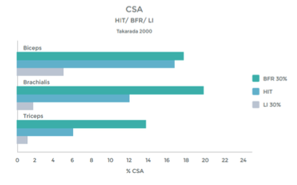
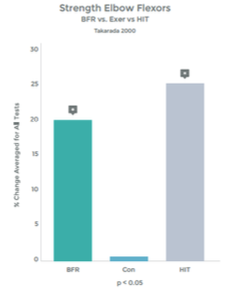
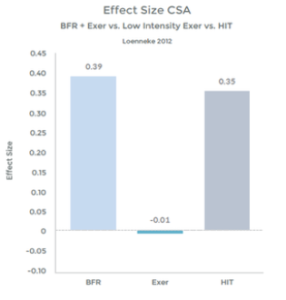
Research comparing the effects of LIT with BFR to High Intensity Training (HIT) helps illustrate the effectiveness even better. This research was done on arm muscles, and the researchers included LIT without BFR as a comparison.1 . See graphs 3 & 4 to the right.

As you can see in the graphs to the right, the changes in strength and hypertrophy of a muscle subjected to BFR training are very similar to the effects seen with High Intensity loading. In fact, the BFR group showed even greater initial changes in muscle ‘cross sectional area’ – Growth!

Many studies have demonstrated significant increases in muscle strength using BFR and exercise, as compared to exercise alone, at low intensity. 2 3 4 5 6 7 8 9 10

Graph 3

Graph 4

Graph 5

Many studies have demonstrated significant increases in muscle hypertrophy using BFR and exercise, as compared to exercise alone, at low intensity 11 12 13 14 15 16 17 18

Many studies have demonstrated that the effects of BFR with LIT are similar to HIT with respect to strength and hypertrophy gains. 19 20 21 22 23 24 25 26

How Does it Work? 

From the available research – which is vast – we are left to believe that multiple mechanisms may be at work. Below is a summary:

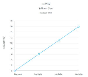
1) Lactate Production

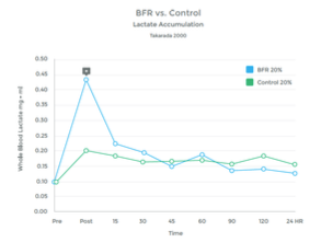
The accumulation of lactic acid in a muscle is a result of ‘anaerobic’ metabolism – work being performed without adequate oxygen. This provides the ‘burn’ of a hard workout to fatigue.

When O2 is plentiful, the muscle utilizes the Kreb Cycle to produce energy needed to perform work. However, when O2 is depleted, the muscle utilizes an alternative mechanism called the Cori Cycle (also known as the Lactic Acid Cycle). Lactate is a by-product of this process.

Graph 6

Graph 7

While the Cori Cycle is typically only activated under high intensity conditions, BFR training appears to be an alternative way to achieve a similar rise in lactate levels.  See graphs 6 & 7 to the right.

Many studies have observed this rise in lactic acid associated with BFR Training. 27 28 29 30 31 32 33
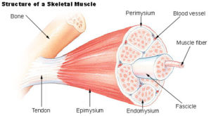
Image 1

Graph 8

2) Improved Muscle Activation

Typically, under low loads, a relatively small amount of the muscle is ‘recruited’ to perform the work. Muscles are made up of hundreds of thousands of individual muscle fibers, which are, themselves, sub-categorized into muscle ‘fascicles’.  (See image to left)

Nerves that innervate the muscle do so by branching out and forming many attachment points. These separate connections are known as ‘motor units’ and they are capable of contracting separate from the other portions of the muscle. A given muscle will have hundreds of motor units. So, with light contraction, a small number of ‘motor units’ are recruited (or turned on), but with heavy effort, more units are enlisted.

Under typical conditions, lactic acid builds up in the muscle only under heavy, fatiguing conditions, thus the body responds to the presence of lactate by up regulating (increasing) motor unit recruitment. The body recruits, not just higher numbers of motor units, but large motor units as well (the ‘units’ with lots of fibers in them, that typically get called to action under high loads)

Studies have shown a clear link between the presence of lactic acid, and an increase in muscle fiber recruitment. (See graph 8 on left)

Anyone who has had knee surgery and couldn’t get the Quad to “wake up” can tell you – this is a big deal!

Multiple studies have confirmed the direct correlation between BFR training and a strong Lactic Acid response. If you doubt it… come try it! You will be sold.

3) Increased activation of ‘Fast Twitch’ Fibers

Under normal conditions, LIT does not recruit these muscle fiber types. ‘Fast Twitch’ muscles are the fibers that fire hard and heavy when a lot of work is needed quickly. They do not rely on oxygen for fuel, as apposed to ‘Slow Twitch’ endurance fibers.  With low loads, these motor units will be recruited only when starved of Oxygen with BFR. 34

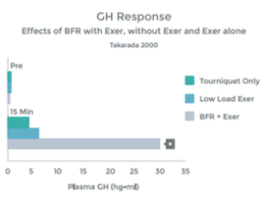
4) Increased Release of Growth Hormone

Growth Hormone (GH) appears to play an important role in muscle recovery. Despite the commonly held belief that GH builds muscle, what it appears to be primarily involved in is the regeneration of Muscle Collagen and Tendon health 35 36 37 38

An accumulation of Lactate – as occurs with HIT and with LIT with BFR – triggers augmented growth hormone release. 39 40 41 42. When the muscle is in use, it stimulates nerve endings within the muscle (known as Type III and Type IV afferent nerves) that communicate with many areas in the body (e.g. your heart and lungs). We know that with heavy use, and the resulting Lactic Acid build up, these nerve stimulate the Pituitary Gland to release Growth Hormone 43. Low Intensity Training with BFR appears to result in a similar response. Please see graph 9.

The results of the study above were replicated by a second study that found, with BFR + LIT, GH levels 290% higher than baseline (resting measurements). This response is 1.7 x than that previously reported to be associated with HIT 44 45

Graph 9

Graph 10

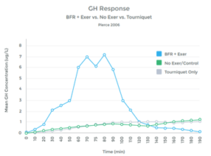
Multiple studies have shown a similar response, as well as a lack of hormonal response when LIT was not combined with BFR. (See graph 10 to left). 46 47 48 49 50 51 52 53 54 55

Thus with BFR training, the body interprets the Lactate build up as an indicator of heavy use and muscle breakdown. The response is to increase the release of GH as a mechanism to repair and regenerate – however, there was significant tissue damage associated with LIT with BFR, thus, the patient ends up with a positive collagen turnover (This is good!)

5) Increased Release of Numerous Hormonal Triggers for Muscle Growth

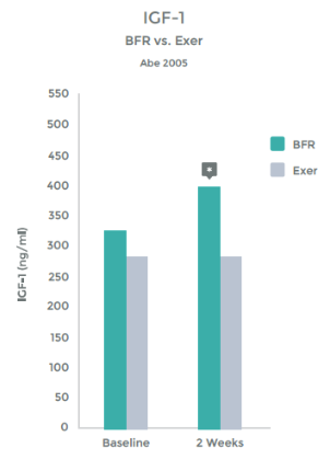
Another hormonal benefit from BFR is that the increase in GH stimulates the release of Insulin like growth factor (IGF-1). IGF-1 does effect muscle hypertrophy (growth), strength gains and some researchers have dubbed it the regulator of muscle mass 56 57 58 59

Multiple studies have shown an increase of IGF-1 levels when BFR is added to exercise.60 61 62

Studies have even shown an increase in IGF-1 levels with LIT + BFR greater than that seen with HIT 63 64 65

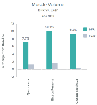
Abe, et al demonstrated this – See graph 11 on right.

In addition, they illustrated the link b/w this hormonal response and muscle hypertrophy. This graph demonstrates the percent change in Cross Sectional Area (CSA), i.e. muscle size with LIT Vs. LIT + BFR. – See graph 12 on right.

So, with BFR training, we trigger the release of GH. This, in turn, activates IGF-1 and muscle stem cells to facilitate muscle hypertrophy 66 – resulting in a bigger, stronger muscle!

Graph 11

Graph 12

Nielson et al did a study looking into the ability of BFR to trigger the IGF-1 pathway and confirmed the increase in numbers of ‘muscle satellite’ cells (essentially stem cells that are capable of building muscle). The extent of this response was dramatic – a 280% increase at mid-training. As a comparison, ~ 30-50% gains are typically seen with HIT 67 68 69

They also examined muscle fiber size and confirmed a 30-40% increase in muscle fiber area during training (most of which was maintained after 7-10 days of ‘de-training’). To put this into perspective 12-16 weeks of heavy resistance training has demonstrated a 15-20% increase in muscle fiber area in untrained men. (See Graphs 13 & 14 on right). 70 71 72

Last, these changes were associated with an increase in strength not demonstrated in the control group. (See Graph 15 on right).

These findings have been repeated 73

Graph 13

Graph 14

Graph 15

6) Increase in MTORC1

MTORC1 serves essentially as a switch to turn on muscle protein synthesis – and thus, build muscle. Bringing a muscle to failure results in an activation of MTORC1. 74  The body says, if you need to use that muscle that hard, let me build it up for you.

While BFR protocols do not use a muscle to lift heavy weight, we due induce a full fatigue and ‘failure’. Studies have shown that BFR + LIT result in increased levels of MTORC1 (See Graph 16 on right).75

Further, this resulted in an increased in muscle protein synthesis – by 46%!

This effect works, even in elderly exercisers 76 – a group known to be resistant to muscle growth 77 78

Multiple studies have confirmed the link between BFR and MTORC1 induced muscle protein synthesis 79 80

Graph 16

In response to the evidence above, Dr. Ronald Meyer, a Physiologist from Michigan State University, stated “the recommendation that hypertrophy requires a load 70% of one repetition maximum might just as well be recast as a recommendation that the training must result in substantial anaerobic metabolism”. Essentially, he is saying that we can no longer hold true to the assertion that muscle only grows in response to heavy loading. Clearly, hypertrophy is a reaction to the metabolic signals associated with anaerobic (Oxygen deprived) conditions. 81

This is ground breaking!

7) Down Regulation of Myostatin

Myostatin is a protein found naturally in our bodies. Its job is to keep muscle development in check. In other words, Increased Myostatin = Decreased Muscle. Decreased Myostatin = Increased Muscle.

The Belgian Blue cattle are a breed deficient in Myostatin. The dog pictured to the left had a genetic mutation that resulted in a Myostatin deficiency.

Myostatin levels are known to lower in response to exercise. The body creates an environment conducive to muscle growth. This typically only occurs in High Intensity Training. 82 83 84 85 86

However, researchers have shown very similar decreases in response to Low Intensity training, when combined with BFR. (See Graph 17 on left). 87

The decrease in Myostatin was actually slightly larger in the BFR group as compared to the HIT group (45% compared to 41%).

Incidentally, in this study, the muscle size improved in both HIT and LIT + BFR groups, but not the LIT alone group. Strength improved 40.1% in the HIT group, 36.2% in the LIT + BFR, and not at all in the LIT alone group.

There have been multiple studies demonstrating the effect BFR training has on Myostatin and its associated effect on muscle hypertrophy and strength gains. 88 89

Image 2

Image 3

Graph 17

Inhibiting Scar Tissue?

Muscle tissue is renown for developing fibrosis (the laying down of ‘scar tissue’) as a result of injury.  Proteins belonging to the TGF-Beta superfamily appear to facilitate this lying down of scar tissue – A good way to ‘bridge the gap’ of injured muscle quickly, but it results in a weaker and less flexible repair. 90 Myostatin is a member of the TGF-Beta family.

Blocking TGF-Beta proteins in animal studies has shown improved regeneration of actual muscle in response to injury. 91

Now, completely blocking TGF-Beta proteins early in the post-injury phase is a bad thing. 92

Some fibrosis is appropriate and important in early healing. However, over development of Fibrotic tissue is not beneficial.

It has been shown that reducing Myostatin levels after an injury, reduces fibrosis (scar tissue) in the muscle. 93  Some researchers have even found that lowering Myostatin levels reverses fibrosis. 94

Blood Flow Restriction Training creates an environment that

  • Ÿ Facilitates tissue healing
  • Ÿ Limits fibrosis (scar tissue formation)
  • Ÿ Enhances muscle growth / hypertrophy

Resulting in

  • Ÿ Decreased (or even eliminated) atrophy for injured or post-operative patients requiring prolonged (> 1 week) time with limited use and/or weight bearing.
  • Ÿ Quicker recovery of tissue heal / structural integrity
  • Ÿ Faster strength gains and return to function

… A Game Changer.

  1. Takarada Y, Takazawa H, Sato Y, Takebayashi S, Tanaka Y, Ishii N. Effects of resistance exercise combined with moderate vascular occlusion on muscular function in humans. J Appl Physiol 88: 2097–2106, 2000. “In the present study, a low-intensity resistance exercise training regimen with moderate vascular occlusion caused a marked muscular hypertrophy, as did a high intensity exercise… Because the effect of HI exercise at 80% 1 RM would represent that close to maximal in inducing muscular hypertrophy, an exercise with occlusion at an intensity even lower than 50% 1 RM is expected to be substantially effective. Such an effectiveness of low intensity exercise disagrees with the established principle for programming resistance exercise, because it has been generally believed that an intensity lower than 65% 1 RM is not useful for gaining muscular size and strength.”
  2. Abe T, Kearns CF, Sato Y. Muscle size and strength are increased following walk training with restricted venous blood flow from the leg muscle, Kaatsu-walk training. J Appl Physiol 100: 1460–1466, 2006.
  3. Abe T, Sakamaki M, Fujita S, Ozaki H, Sugaya M, Sato Y, Nakajima
T. Effects of low-intensity walk training with restricted leg blood flow on muscle strength and aerobic capacity in older adults. J Geriatr Phys Ther 33: 34–40, 2010.
  4. Abe T, Sato Y, Inoue K, Midorikawa T, Yasuda T, Kearns CF, Koizumi K, Ishii N. Muscle size and IGF-1 increased after two weeks of low-intensity “Kaatsu” resistance training (Abstract). Med Sci Sports Exerc 36: S353, 2004.
  5. Evans C, Vance S, Brown M. Short-term resistance training with blood flow restriction enhances microvascular filtration capacity of human calf muscles. J Sports Sci 28: 999–1007, 2010.
  6. Drummond MJ, Fujita S, Takash A, Dreyer HC, Volpi E, Rasmussen BB. Human muscle gene expression following resistance exercise and blood flow restriction. Med Sci Sports Exerc 40: 691–698, 2008.
  7. Madarame H, Neya M, Ochi E, Nakazato K, Sato Y, Ishii N. Crosstransfer effects of resistance training with blood flow restriction. Med Sci Sports Exerc 40: 258– 263, 2008.
  8. Patterson SD, Ferguson RA. Increase in calf post-occlusive blood flow and strength following short-term resistance exercise training with blood Flow restriction in young women. Eur J Appl Physiol 108: 1025–1033, 2010.
  9. Takarada Y, Takazawa H, Sato Y, Takebayashi S, Tanaka Y, Ishii N. Effects of resistance exercise combined with moderate vascular occlusion on muscular function in humans. J Appl Physiol 88: 2097–2106, 2000.
  10. Yasuda T, Fujita S, Ogasawara R, Sato Y, Abe T. Effects of low intensity bench press training with restricted arm muscle blood flow on chest muscle hypertrophy: a pilot study. Clin Physiol Funct Imaging 30: 338–343, 2010.
  11. Abe T, Kearns CF, Sato Y. Muscle size and strength are increased following walk training with restricted venous blood flow from the leg muscle, Kaatsu-walk training. J Appl Physiol 100: 1460–1466, 2006.
  12. Abe T, Sakamaki M, Fujita S, Ozaki H, Sugaya M, Sato Y, Nakajima
T. Effects of low-intensity walk training with restricted leg blood flow on muscle strength and aerobic capacity in older adults. J Geriatr Phys Ther 33: 34–40, 2010.
  13. Abe T, Sato Y, Inoue K, Midorikawa T, Yasuda T, Kearns CF, Koizumi K, Ishii N. Muscle size and IGF-1 increased after two weeks of low-intensity “Kaatsu” resistance training (Abstract). Med Sci Sports Exerc 36: S353, 2004.
  14. Madarame H, Neya M, Ochi E, Nakazato K, Sato Y, Ishii N. Cross transfer effects of resistance training with blood flow restriction. Med Sci Sports Exerc 40: 258–263, 2008.
  15. Martin-Hernandez, J., Marin, P. J., Menendez, H., Loenneke, J. P., Coelho -e-Silva, M. J., Garcia-Lopez, D., & Herrero, A. J. (2013). Changes in muscle architecture induced by low load blood flow restricted training. Acta Physiol Hung, 100(4), 411-418.
  16. Takarada Y, Takazawa H, Sato Y, Takebayashi S, Tanaka Y, Ishii N. Effects of resistance exercise combined with moderate vascular occlusion on muscular function in humans. J Appl Physiol 88: 2097–2106, 2000.
  17. Yasuda, T., Fukumura, K., Uchida, Y., Koshi, H., Iida, H., Masamune, K., . . . Nakajima, T. (2014). Effects of Low-Load, Elastic Band Resistance Training Combined With Blood Flow Restriction on Muscle Size and Arterial Stiffness in Older Adults. J Gerontol A Biol Sci Med Sci.
  18. Wilson, J. M., Lowery, R. P., Joy, J. M., Loenneke, J. P., & Naimo, M. A. (2013). Practical blood flow restriction training increases acute determinants of hypertrophy without increasing indices of muscle damage. J Strength Cond Res, 27(11), 3068-3075
  19. Martin-Hernandez, J., P. J. Marin, H. Menendez, C. Ferrero, J. P. Loenneke, and A. J. Herrero. 2013. Muscular adaptations after two different volumes of blood flow- restricted training. Scand. J. Med. Sci. Sports 23:e114–e120.
  20. Vechin, F. C., C. A. Libardi, M. S. Conceicao, F. R. Damas, M. E. Lixandrao, R. P. Berton, et al. 2015. Comparisons between low-intensity resistance training with blood flow restriction and high-intensity resistance training on quadriceps muscle mass and strength in elderly. J. Strength Cond. Res. 29:1071–1076.
  21. Laurentino, G. C., Ugrinowitsch, C., Roschel, H., Aoki, M. S., Soares, A. G., Neves, M., Jr., Tricoli, V. (2012). Strength training with blood flow restriction diminishes myostatin gene expression. Med Sci Sports Exerc, 44(3), 406-412.
  22. Lowery, Ryan P., et al. “Practical blood flow restriction training increases muscle hypertrophy during a periodized resistance training programme.“Clinical physiology and functional imaging 34.4 (2014): 317-321.
  23. Takarada, Y., Takazawa, H., Sato, Y., Takebayashi, S., Tanaka, Y., & Ishii, N. (2000). Effects of resistance exercise combined with moderate vascular occlusion on muscular function in humans. J Appl Physiol (1985), 88(6), 2097- 2106.
  24. Clark, B. C., et al. (2011). “Relative safety of 4 weeks of blood flow-restricted resistance exercise in young, healthy adults.” Scand J Med Sci Sports 21(5): 653-662.
  25. Kubo K, Komuro T, Ishiguro N, et al.. Effects of low-load resistance training with vascular occlusion on the mechanical properties of muscle and tendon. J Appl Biomech. 2006; 22 (2): 112–9.
  26. Karabulut M, Abe T, Sato Y, Bemben MG. The effects of low-intensity resistance training with vascular restriction on leg muscle strength in older men. Eur J Appl Physiol. 2010; 108 (1): 147–55.
  27. Laurentino G, Ugrinowitsch C, Aihara AY, et al. Effects of strength training and vascular occlusion. Int J Sports Med. 2008; 29(8): 664–7.
  28. Loenneke JP, Wilson JM, Marin PJ, et al. Low intensity blood flow restriction training: a meta- analysis. Eur J Appl Physiol.2012; 112(5):1849–59.
  29. Loenneke, J. P., Kearney, M. L., Thrower, A. D., Collins, S., & Pujol, T. J. (2010). The acute response of practical occlusion in the knee extensors. J Strength Cond Res, 24(10), 2831-2834.
  30. Poton, R., & Polito, M. D. (2014). Hemodynamic response to resistance exercise with and without blood flow restriction in healthy subjects. Clin Physiol Funct Imaging. doi: 10.1111/cpf.12218
  31. Scott, B. R., Loenneke, J. P., Slattery, K. M., & Dascombe, B. J. (2014). Exercise with Blood Flow Restriction: An Updated Evidence-Based Approach for Enhanced Muscular Development. Sports Med. doi: 10.1007/s40279-014-0288-1
  32. Sundberg, C. J. (1994). Exercise and training during graded leg ischaemia in healthy man with special reference to effects on skeletal muscle. Acta Physiol Scand Suppl, 615, 1-50
  33. Takarada, Y., Takazawa, H., Sato, Y., Takebayashi, S., Tanaka, Y., & Ishii, N. (2000). Effects of resistance exercise combined with moderate vascular occlusion on muscular function in humans. J Appl Physiol (1985), 88(6), 2097-2106.
  34. Wilson JM, Lowery RP, Joy JM, Loenneke JP, Naimo MA. Practical blood flow restriction training increases acute determinants of hypertrophy without increasing indices of muscle damage. J Strength Cond Res 2013;27:3068–75.
  35. Doessing S, Heinemeier KM, Holm L, et al. Growth hormone stimulates the collagen synthesis in human tendon and skeletal muscle without affecting myofibrillar protein synthesis. J Physiol. 2010;588(Pt 2):341–51.
  36. Kurtz CA, Loebig TG, Anderson DD, DeMeo PJ, Campbell PG. Insulin-like growth factor I accelerates functional recovery from Achilles tendon injury in a rat model. Am J Sports Med 1999: 27(3): 363– 369.
  37. Baroncelli GI, Bertelloni S, Ceccarelli C, Cupelli D, Saggese G. Dynamics of bone turnover in children with GH deficiency treated with GH until final height. Eur J Endocrinol 2000: 142(6): 549–556.
  38. Boesen, A. P., Dideriksen, K., Couppe, C., Magnusson, S. P., Schjerling, P., Boesen, M., Langberg, H (2014). Effect of growth hormone on aging connective tissue in muscle and tendon: gene expression, morphology, and function following immobilization and rehabilitation. J Appl Physiol (1985), 116(2), 192-203. doi: 10.1152/japplphysiol.01077.2013
  39. Gordon, S.E., Kraemer, W.J., Vos, N.H., Lynch, J.M., & Knuttgen, H.G. (1994). Effect of acid-base balance on the growth hormone response to acute high-intensity cycle exercise. Journal of Applied Physiology, 76, 821–829.
  40. Goto, K., Ishii, N., Kizuka, T., & Takamatsu, K. (2005). The impact of metabolic stress on hormonal responses and muscular adaptations. Med Sci Sports Exerc, 37(6) 955-963.
  41. Takarada Y, Nakamura Y, Aruga S, Onda T, Miyazaki S, Ishii N. Rapid increase in plasma growth hormone after low-intensity resistance exercise with vascular occlusion. J Appl Physiol (1985). 2000; 88:61-65.
  42. Godfrey RJ, Whyte GP, Buckley J, Quinlivan R. The role of lactate in the exercise-induced human growth hormone response: evidence from McArdle disease. Br J Sports Med. 2009;43:521-525.
  43. Gosselink, K. L., R. E. Grindeland, R. R. Roy,H. Zhong, A. J. Bigbee, E. J. Grossman, and V. R. Edgerton. Skeletal muscle afferent regulation of bioassayable growth hormone in the rat pituitary. J. Appl. Physiol. 84: 1425– 1430, 1998.
  44. Takarada Y, Nakamura Y, Aruga S, Onda T, Miyazaki S, Ishii N. Rapid increase in plasma growth hormone after low-intensity resistance exercise with vascular occlusion. J Appl Physiol (1985). 2000; 88:61-65.
  45. Kraemer, W. J., Marchitelli, L., Gordon, S. E., Harman, E., Dziados, J. E., Mello, R., Fleck, S. J. (1990). Hormonal and growth factor responses to heavy resistance exercise protocols. J Appl Physiol (1985), 69(4), 1442-1450.
  46. Pierce, J. R., Clark, B. C., Ploutz-Snyder, L. L., & Kanaley, J. A. (2006). Growth hormone and muscle function responses to skeletal muscle ischemia. J Appl Physiol (1985), 101(6), 1588-1595. doi:
  47. Manini, T. M., Yarrow, J. F., Buford, T. W., Clark, B. C., Conover, C. F., & Borst, S. E. (2012). Growth hormone responses to acute resistance exercise with vascular restriction in young and old men. Growth Horm IGF Res, 22(5), 167-172.
  48. Kraemer, W. J., Marchitelli, L., Gordon, S. E., Harman, E., Dziados, J. E., Mello, R., Fleck, S. J. (1990). Hormonal and growth factor responses to heavy resistance exercise protocols. J Appl Physiol (1985), 69(4), 1442-1450.
  49. Kraemer, W. J., S. E. Gordon, S. J. Fleck, L. J. Marchitelli, R. Mello, J. E. Dziados, K. Friedl, E. Harman, C. Maresh, and A. C. Fry. Endogenous anabolic hormonal and growth factor responses to heavy resistance exercise in males and females. Int. J. Sports Med. 12: 228–235, 1991.
  50. Fujita S, Abe T, Drummond MJ, et al. Blood flow restriction during low-intensity resistance exercise increases S6K1 phosphorylation and muscle protein synthesis. J Appl Physiol. 2007; 103:903– 910.
  51. Madarame H, Neya M, Ochi E, Nakazato K, Sato Y, Ishii N. Cross-transfer
effects of resistance training with blood flow restriction. Med Sci Sports Exerc. 2008; 40:258–263
  52. Madarame H, Sasaki K, Ishii N. Endocrine responses to upper- and lower-limb resistance exercises with blood flow restriction. Acta Physiol Hung. 2010; 97:192–200.
  53. Yasuda T, Fujita S, Ogasawara R, Sato Y, Abe T. Effects of low-intensity bench press training with restricted arm muscle blood flow on chest muscle hypertrophy: a pilot study. Clin Physiol Funct Imaging. 2010; 30: 338–343.
  54. Reeves GV, Kraemer RR, Hollander DB, et al. Comparison of hormone responses following light resistance exercise with partial vascular occlusion and moderately difficult resistance exercise without occlusion. J Appl Physiol. 2006; 101:1616–1622.
  55. Takano H, Morita T, Iida H, et al. Hemodynamic and hormonal responses to a short- term low intensity resistance exercise with the reduction of muscle blood flow. Eur J Appl Physiol. 2005; 95:65–73.
  56. Haddad F, Adams GR. Inhibition of MAP/ERK kinase prevents IGF-I-induced hypertrophy in rat muscles. J Appl Physiol. 2004;96(1):203–10.
  57. Stewart CE, Pell JM. Point:Counterpoint: IGF is/is not the major physiological regulator of muscle mass. Point: IGF is the major physiological regulator of muscle mass. J Appl Physiol. 2010;108(6):1820,1; discussion 1823-4; author reply 1832.
  58. Hameed M, Lange KH, Andersen JL, et al. The effect of recombinant human growth hormone and resistance training on IGF-I mRNA expression in the muscles of elderly men. J Physiol. 2004;555(Pt 1):231–40.
  59. Kostek MC, Delmonico MJ, Reichel JB, et al. Muscle strength response to strength training is influenced by insulin-like growth factor 1 genotype in older adults. J Appl Physiol. 2005;98(6): 2147–54
  60. Abe T, Yasuda T, Midorikawa T, et al. Skeletal muscle size and circulating IGF-1 are increased after two weeks of twice daily KAATSU resistance training. Int J Kaatsu Train Res. 2005;1: 6–12.
  61. Takano H, Morita T, Iida H, et al. Hemodynamic and hormonal responses to a short-term low-intensity resistance exercise with the reduction of muscle blood flow. Eur J Appl Physiol. 2005;95(1):65– 73.
  62. Fujita S, Abe T, Drummond MJ, et al. Blood flow restriction during low-intensity resistance exercise increases S6K1 phosphorylation and muscle protein synthesis. J Appl Physiol. 2007;103(3):903– 10.
  63. Kraemer WJ, Marchitelli L, Gordon SE, et al. Hormonal and growth factor responses to heavy resistance exercise protocols. J Appl Physiol. 1990;69(4):1442–50.
  64. Kraemer WJ, Gordon SE, Fleck SJ, et al. Endogenous anabolic hormonal and growth factor responses to heavy resistance exercise in males and females. Int J Sports Med. 1991;12(2): 228–35.
  65. Rubin MR, Kraemer WJ, Maresh CM, et al. High-affinity growth hormone binding protein and acute heavy resistance exercise. Med Sci Sports Exerc. 2005;37(3):395–403.
  66. Nielsen, J. L., Aagaard, P., Bech, R. D., Nygaard, T., Hvid, L. G., Wernbom, M., . Frandsen, U. (2012). Proliferation of myogenic stem cells in human skeletal muscle in response to low-load resistance training with blood flow restriction. J Physiol, 590(Pt 17), 4351-4361. 773–782.
  67. Kadi F & Thornell LE (2000). Concomitant increases in myonuclear and satellite cell content in female trapezius muscle following strength training. Histochem Cell Biol 113, 99–103.
  68. Kadi F, Schjerling P, Andersen LL, Charifi N, Madsen JL, Christensen LR & Andersen JL (2004). The effects of heavy resistance training and detraining on satellite cells in human skeletal muscles. J Physiol 558, 1005–1012.
  69. Olsen S, Aagaard P, Kadi F, Tufekovic G, Verney J, Olesen JL, Suetta C & Kjaer M (2006). Creatine supplementation augments the increase in satellite cell and myonuclei number in human skeletal muscle induced by strength training. J Physiol 573, 525–534.
  70. Aagaard P, Andersen JL, Dyhre-Poulsen P, Leffers AM,Wagner A, Magnusson SP, Halkjaer-Kristensen J & Simonsen EB (2001). A mechanism for increased contractile strength of human pennate muscle in response to strength training: changes in muscle architecture. J Physiol 534, 613–623.
  71. Kadi F, Schjerling P, Andersen LL, Charifi N, Madsen JL, Christensen LR & Andersen JL (2004). The effects of heavy resistance training and detraining on satellite cells in human skeletal muscles. J Physiol 558, 1005–1012.
  72. Olsen S, Aagaard P, Kadi F, Tufekovic G, Verney J, Olesen JL, Suetta C & Kjaer M (2006). Creatine supplementation augments the increase in satellite cell and myonuclei number in human skeletal muscle induced by strength training. J Physiol 573, 525–534.
  73. Wernbom, M., Apro, W., Paulsen, G., Nilsen, T. S., Blomstrand, E., & Raastad, T. (2013). Acute low-load resistance exercise with and without blood flow restriction increased protein signalling and number of satellite cells in human skeletal muscle. Eur J Appl Physiol, 113(12), 2953-2965.
  74. Mitchell CJ, Churchward-Venne TA, West DWD, et al. Resistance exercise load does not determine  training-mediated hypertrophic gains in young men. Journal of Applied Physiology.  2012;113(1):71-77. doi:10.1152/japplphysiol.00307.2012.
  75. Fujita S, Abe T, Drummond MJ, et al. Blood flow restriction during low-intensity resistance exercise increases S6K1 phosphorylation and muscle protein synthesis. J Appl Physiol. 2007;103(3):903– 10.
  76. Fry, C. S., Glynn, E. L., Drummond, M. J., Timmerman, K. L., Fujita, S., Abe, T., Rasmussen, B. B. (2010). Blood flow restriction exercise stimulates mTORC1 signaling and muscle protein synthesis in older men. J Appl Physiol (1985), 108(5), 1199-1209.
  77. Sheffield-Moore M, Paddon-Jones D, Sanford AP, Rosenblatt JI, Matlock AG, Cree MG, Wolfe RR. Mixed  muscle and hepatic derived plasma protein metabolism is differentially regulated in older and  younger men following resistance exercise. Am J Physiol Endocrinol Metab288: E922–E929,  2005.
  78. Kumar V, Selby A, Rankin D, Patel R, Atherton P, Hildebrandt W, Williams J, Smith K, Seynnes O,  Hiscock N, Rennie MJ. Age-related differences in the dose-response relationship of muscle  protein synthesis to resistance exercise in young and old men. J Physiol587: 211–217, 2009.
  79. Gundermann, D. M., Walker, D. K., Reidy, P. T., Borack, M. S., Dickinson, J. M., Volpi, E., & Rasmussen, B. B. (2014). Activation of mTORC1 signaling and protein synthesis in human muscle following blood flow restriction exercise is inhibited by rapamycin. Am J Physiol Endocrinol Metab, 306(10), E1198-1204.
  80. Drummond MJ, Fry CS, Glynn EL, Dreyer HC, Dhanani S, Timmerman KL, Volpi E, Rasmussen BB.  Rapamycin administration in humans blocks the contraction-induced increase in skeletal muscle  protein synthesis. J Physiol 587: 1535–1546, 2009.
  81. Meyer, R. A. (2006). Does blood flow restriction enhance hypertrophic signaling in skeletal muscle? J Appl Physiol (1985), 100(5), 1443-1444
  82. Roth SM, Martel GF, Ferrell RE, Metter EJ, Hurley BF, Rogers MA. Myostatin gene expression is reduced in humans with heavy-resistance strength training: a brief communication. Exp Biol Med(Maywood). 2003;228(6):706–9.
  83. Forbes D, Jackman M, Bishop A, Thomas M, Kambadur R, Sharma M. Myostatin 
auto-regulates its expression by feedback loop through Smad7 dependent mechanism. J Cell Physiol. 2006; 206(1). 264–72.
  84. Hill JJ, Qiu Y, Hewick RM, Wolfman NM. Regulation of myostatin in vivo by growth and differentiation factor–associated serum protein-1: a novel protein with protease inhibitor and follistatin domains. Mol Endocrinol. 2003;17(6):1144–54.
  85. Saremi A, Gharakhanloo R, Sharghi S, Gharaati MR, Larijani B, Omidfar K. Effects of oral creatine and resistance training on serum myostatin and GASP-1. Mol Cell Endocrinol. 2010;317(1–2):25–30.
  86. Willoughby DS. Effects of heavy resistance training on myostatin mRNA and protein expression. Med Sci Sports Exerc. 2004;36(4):574–82.
  87. Laurentino, G. C., Ugrinowitsch, C., Roschel, H., Aoki, M. S., Soares, A. G., Neves, M., Jr., Tricoli, V. (2012). Strength training with blood flow restriction diminishes myostatin gene expression. Med Sci Sports Exerc, 44(3), 406-412.
  88. Gualano, B., Neves, M., Jr., Lima, F. R., Pinto, A. L., Laurentino, G., Borges, C. Ugrinowitsch, C. (2010). Resistance training with vascular occlusion in inclusion body myositis: a case study. Med Sci Sports Exerc, 42(2), 250-254.
  89. Santos, A. R., Neves, M. T., Jr., Gualano, B., Laurentino, G. C., Lancha, A. H., Jr., Ugrinowitsch, C., Aoki, M. S. (2014). Blood flow restricted resistance training attenuates myostatin gene expression in a patient with inclusion body myositis. Biol Sport, 31(2), 121-124.
  90. Branton, M. H., & Kopp, J. B. (1999). TGF-beta and fibrosis. Microbes Infect, 1(15), 1349-1365.
  91. Terada, S., Ota, S., Kobayashi, M., Kobayashi, T., Mifune, Y., Takayama, K., Huard, J. (2013). Use of an antifibrotic agent improves the effect of platelet-rich plasma on muscle healing after injury. J Bone Joint Surg Am, 95(11), 980-988.
  92. Gumucio, J. P., Flood, M. D., Phan, A. C., Brooks, S. V., & Mendias, C. L. (2013). Targeted inhibition of TGF-beta results in an initial improvement but long-term deficit in force production after contraction-induced skeletal muscle injury. J Appl Physiol (1985), 115(4), 539-545.
  93. Wagner, K. R. (2005). Muscle regeneration through myostatin inhibition. Curr. Opin. Rheumatol. 17, 720- 724.
  94. Bo Li Z1, Zhang J, Wagner KR. Inhibition of myostatin reverses muscle fibrosis through apoptosis. J Cell Sci. 2012 Sep 1;125(Pt 17):3957-65. doi: 10.1242/jcs.090365. Epub 2012 Jun 8.